Personalizing Stem Cell Therapy for Parkinson’s Disease
October 6, 2017
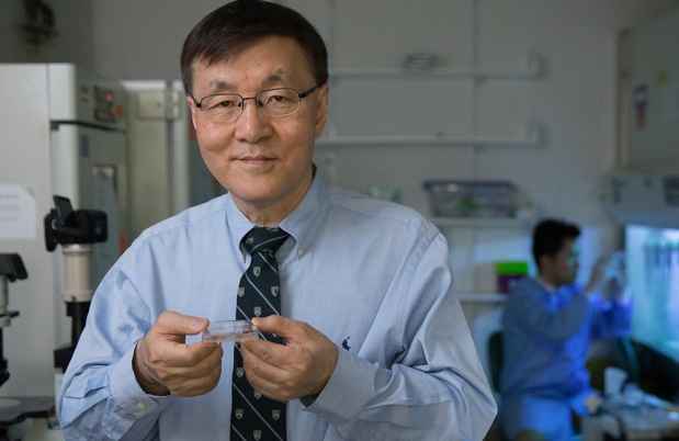
In the United States alone, as many as one million people have Parkinson’s disease (PD), with approximately 60,000 cases being diagnosed each year, according to the Parkinson’s Disease Foundation. Kwang-Soo Kim, PhD, director of the Molecular Neurobiology Laboratory at McLean Hospital and professor of psychiatry at Harvard Medical School, is committed to finding a way to lower those numbers.
Kim’s team is at the forefront of investigating personalized, stem cell-based therapy for PD. He has developed a novel, highly efficient, and safe way to reprogram patients’ own cells to become functioning midbrain dopamine cells—the cells whose loss primarily causes the disease. These cells, which produce the neurotransmitter dopamine, occupy a specific area in the brain’s substantia nigra and project into the dorsal striatum, forming the so-called nigrastriatal pathway. This is the major neuronal pathway controlling human movement, with its disruption leading to the tremors, stiffness, gait changes, and difficulty initiating movements that characterize PD.
Kim’s method overcomes the limitations of other stem cell therapies for PD. It uses patients’ own cells, derived from their skin and transformed into induced pluripotent stem (iPS) cells—cells that behave like embryonic stem cells in that they can develop into practically any cell type. The strategy eliminates not just the possibility of immune rejection but also the ethical issues tied to embryonic stem cell research. It relies on non-viral, footprint-free methods to reprogram the skin cells (such as protein-based or plasmid-based methods) rather than the standard viral delivery system that carries the risk of disrupting patients’ chromosomal DNA and canceling out tumor suppressor genes or activating cancer-causing ones.

Finally, his method removes any remaining “undifferentiated” cells—those that have not yet become a specific cell type—before transplantation, thereby prohibiting tumor formation. Kim’s group added this important step when they discovered that several small molecules, including the natural compound quercetin, selectively kill undifferentiated stem cells. They do so by targeting the survivin gene (BIRC5), which is specifically abundant in undifferentiated cells.
“In terms of cell therapy, the most important criterion is safety,” said Kim. “We now have a very robust, optimized protocol for iPS cell-based autologous cell therapy. Many studies have shown that tumor formation after transplantation is directly proportional to the amount of remaining undifferentiated cells. Our method removes them completely. And by using a non-viral method to reprogram the iPS cells, we can maintain the cells’ genetic integrity.”
The Role of Dopamine in Parkinson’s Disease
Kim has been studying the molecular biology of dopamine neuronal systems for more than 25 years, 18 of them at McLean. His research at the hospital concentrates on the development and maintenance of the neurons themselves. “What makes a dopamine neuron become a dopamine neuron?” he said, describing it as the burning question he wanted to address. “What goes wrong with dopamine neurons in the case of disease?”
Turning to stem cell research to answer these questions was a natural progression. Stem cells are essentially a tabula rasa: they can give rise, through the process of differentiation, to any kind of specialized cell, for example, a heart, liver, or muscle cell, depending on how the stem cell’s genes interact with the physical and chemical conditions in its environment.
Kim set out to find what would turn stem cells into the specific dopamine cells degenerating in PD—midbrain dopamine (mDA) neurons in the substantia nigra. He initially used mouse embryonic stem cells to generate mDA neurons and transplanted them into the brains of mice or rats engineered to have traits of PD.
“We could see that this transplantation had the potential to rescue the motor function defect in the animals,” said Kim. “But at the same time, there were some very significant limitations in that some of the animals developed tumors, and differentiation to mDA cells was not very efficient.” In 2009, he shifted his attention to human iPS cells generated from human skin cells. Using a direct protein method to deliver the genes that would trigger the transformation eliminated the risk of disrupting the cells’ chromosomal DNA, but the process was slow, and the produced iPS cells varied in their ability to differentiate into mDA neurons.
Recently, Kim and his colleagues discovered a fundamental mechanism underlying the change in metabolic properties that accompanies the skin cells’ reprogramming. The finding enabled them to develop a novel reprogramming method, for which they are awaiting a patent.
“Based on our new method, we can make iPS cells with much greater efficiency, and they show much less variation,” Kim said. “They are almost standardized in their ability to differentiate into mDA neurons.”
Optimizing the Differentiation Protocol
Differentiating iPS cells into particular cell types is tricky, but Kim’s lab has gone a long way toward doing so, spending countless hours bent over petri dishes investigating which signals transform them into functional mDA neurons. One of these differentiation technologies was recently licensed to the stem cell company Cellular Dynamics, based in Madison, Wisconsin, and the protocol itself is patent pending.
“My lab is continuing to optimize the differentiation protocol, because if you think about brain development, there are maybe tens or even hundreds of different signals taking place in a temporal-specific manner, in a gradient-specific manner, in a combinatorial manner to drive specific neuronal lineages,” said Kim. “We now have an effective protocol that can make large proportions of mDA neurons.”
“My hope is that in the not too distant future we will be ready to run a human clinical trial.”– Kwang-Soo, Kim, PhD
The current challenge is to test the human mDA neurons in an animal model to determine the best stage at which to transplant them and how many to transplant to reverse Parkinson’s effects. For that, Kim and his colleagues are using the popular 6-OHDA lesion rat model. The researchers inject the chemical 6-OHDA, which kills dopamine neurons, into one side of the nigrastriatal pathway, causing a lesion and stopping dopamine function. Behaviorally, upon administration of apomorphine, the rat will rotate to the lesion side, as movements will now be driven by the intact side. They then inject the new mDA neurons into the lesion side, wait three to four months for the transplant to “take” and new neurons to grow, and make careful computer-generated measurements of the rat’s rotation recovery. Later they sacrifice the animals to analyze the extent of the neuronal growth in their brains and how that growth relates to the elimination of symptoms.
“With all this analysis, we can come up with the most accurate calculations for real transplant conditions,” said Kim. “My hope is that in the not too distant future we will be ready to run a human clinical trial.”
Media Requests
Journalist or member of the media? We are available 24/7 for media requests.